|
Identity
For a variety of
reasons, aquarists often want to attach names of one sort
or another to the animals in their systems. Generally, the
way this is done is that the hobbyist goes to some reference
and finds a picture of an organism that "looks"
like the one in his system and, "Presto! The organism
has been identified." While this approach will work moderately
well with fishes, some snails, many echinoderms and some shrimps,
it doesn't work worth a squat with the sponges.
For such a visually comparative methodology to be successfully
applied, the organisms within the species in question must
have little variation in shape or color from one individual
to the next and there must be few other species with similar
morphologies. We all know that organisms within a species
vary somewhat in morphological characteristics such as color
and shape. The example of this type of variability with which
most of us are probably familiaris our own species. It is
obvious that not all humans look alike, but experience has
shown that all human shapes and sizes can successfully interbreed.
At the same time, it is also evident that there is little
variation in appearance across the whole human species. This
follows from the type of animal that we are; all vertebrates
have an adult shape that is relatively consistent across their
species. Most animals are similar in this regard; within a
species, one individual looks pretty much like every other
individual.
A number of animal groups, however, do things very differently.
Growth in these animals doesn't lead to a defined adult shape
or morphology. While their growth is always governed by some
genetically determined rules, these rules don't necessarily
lead to a defined adult size or shape. Put another way, even
though there will be some structural similarities, no two
animals in the species will have the same shape. Within such
marine species, the growth form is often determined by environmental
variables such as water currents or light, and the final adult
shape is due probably as much to the environment as it is
to the animal's inherited characteristics. If several species
have the same basic genetic rules for growth, individuals
from those species will, for all intents and purposes, look
identical if they are grown under similar environmental conditions.
Sponges are animals that grow this way, and within every geographic
region many species often appear, to the casual observer,
to be identical. The problem is compounded, however, when
comparing species from many different geographic regions.
Unless the potential identifier has a great degree of familiarity
with a specific region, it becomes well nigh impossible to
identify such animals.
|
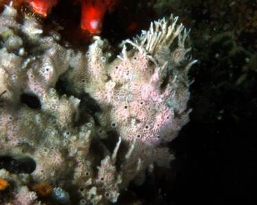
Figure 1. Sigmadocia sp. This temperate
sponge was growing in the lee of a rock and shows the
effects of changing currents on colonial shape. As the
colony grew up into an area of vigorous currents (to
the upper right), its morphology changed from a massive
encrusting form to one with thin linear branches. This
sponge was collected and identified in my laboratory.
Identification by appearance alone was impossible.
|
 |
Sponge species, as whole, are impossible to identify by casual
comparison to some photograph or illustration. It is possible
to identify the animals, of course; it just usually cannot
be done by simply comparing their gross appearance. That having
been said, every geographic region has a number of sponges
that are easily identifiable in this manner. Unfortunately,
with few exceptions, these easily identifiable animals are
not generally the ones that are able to be kept in marine
aquaria. Within the sponge groups that I discussed last
month, probably the ones with the most consistent apparent
morphologies are the hexactinellids (the glass sponges) and
the calcareous sponges. In the most diverse, and most common,
sponge group, the demosponges, most species are impossible
to identify by comparison with photographs.
Obviously, the plasticity of shape seen in most demosponges
leads to the obvious question, "If these animals cannot
be identified to species simply by looking at them, how are
they identified?" The answer is relatively straightforward,
actually, as is the identification process. Some of the animal's
internal structures must be compared to the corresponding
structures found in other similar animals. The earliest taxonomists
recognized the variability of sponge shape as an impediment
to these animals' easy identification and characterization,
and it was realized early on that other types of criteria
would be necessary to reliably distinguish between sponges.
In addition to the variability of sponge shape, another problem
was the basic simplicity of sponges. Sponges simply have few
structures on their bodies that can be used for identification.
Animals such as fishes have all sorts of external characteristics
that can be used to distinguish between species. They can
vary in color, body shape, fin shape, fin length, presence
or absence of scales, the shapes of scales, the presence of
spines, spine shape, and so on ad infinitum. In contrast,
sponges are blobs with holes of several different sizes in
them. The paucity of structures makes them difficult to easily
characterize - and this has lead to many problems with their
taxonomy. About the only consistent structures in most sponges
are their spicules.
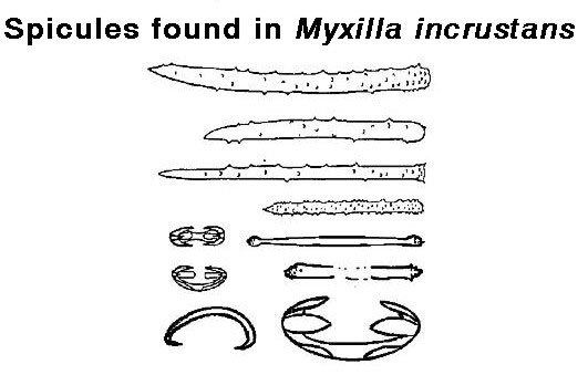
Consequently, the sizes, shapes, and relative abundances
of these microscopic skeletal structures are the structures
used to distinguish between various sponge species.
By the way, hobbyists are not the only people who have problems
identifying sponges; many sponge experts have had problems
as well, and that has lead to everybody subsequent to them
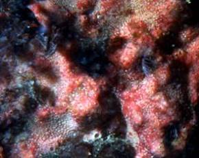
having problems. As an example of this confusion, consider
the common intertidal sponge Ophlitaspongia pennata
found along the Pacific Coast of North America. This species
is quite abundant, and is commonly found in the lower intertidal
zone. It was originally described by Lambe in 1893. He described
the spicular array that he considered to be diagnostic of
the species. He also described it as having a specific and
characteristic morphology in which the sponge's water channels
were quite evident. Few shallow water sponges along the Pacific
Coast have a characteristic morphology, but for several decades
it was thought that Ophlitaspongia was one of them.
Virtually all examples of this sponge found in shallow water
show the characteristic surface with the visible water channel
system. Ophlitaspongia is also notable for one other
reason; it is a brilliant red, and that coloration makes it
strikingly obvious. It is the only red sponge common throughout
the region. Although there was another red sponge, it was
considered to be rare and was collected from deeper waters.
This other sponge was obviously not Ophlitaspongia
as it had 1) a different appearance, 2) a different array
of internal spicules and 3) was found in a different habitat.
The rare red sponge was essentially impossible to identify
with the available literature. It had a different spicule
array than what was known from the area's other sponges. Because
of the differences from Ophlitaspongia, it appeared to be
a species that was "new to science." However, Ophlitaspongia
pennata in the intertidal zone is almost always found
with a small nudibranch, Rostanga pulchra, grazing
on it. This little nudibranch is rarely found in deeper water,
but that wasn't known at the time. It wasn't until some students
started to examine the relationship between this particular
nudibranch and its prey that it became evident that the "characteristic"
appearance of the sponge was the result of having been partially
eaten by the snail. Upon removal of the nudibranch from the
sponge, the sponge's shape and spicule arrangement changed
quite drastically, and it turned out that the rare deep-water
red sponge was simply Ophlitaspongia growing in the
absence of predation (Bakus, 1966; Anderson, 1971).
|
|
 |
|
Figure 2. Ophlitaspongia pennata.
Left: A shallow water specimen with the nudibranch,
Rostanga pulchra, commonly found on it. The nudibranch
is about 1 cm (0.4 in) long. Note the visible water
channel canals caused by the nudibranch's grazing of
the sponge's surface. Right: A deep-water specimen
lacking the nudibranch and showing no signs of grazing.
Note the difference in surface shape and texture. The
white dots on the sponge are spirorbid worms similar
to those commonly found in marine aquaria.
|
Step 1. To What Major Group Do the Sponges Belong?
The first thing anyone identifying
sponges has to do is to identify the animal to its basic group.
In other words, "Is the sponge a hexactinellid, a demosponge
or a calcareous sponge?" Hexactinellids are rare in shallow
water, and are almost never seen on coral reefs, so most hobbyists
can eliminate that choice immediately; nevertheless, in the
unlikely event that one could turn up, I have included them
in this discussion.
To determine a sponge's major group, first place a small
piece of it in a small glass container (a vial or very small
jar works well). Add enough chlorine bleach to cover it, then
shake it well and let it stand for a while. When the sample
quits bubbling, use a pipette to remove the fluid, being careful
not remove any of the residue at the bottom. If the piece
dissolves completely and no residue remains, the sponge is
a demosponge. Some demosponges (such as bath sponges) will
leave no residue at all after the bleach treatment.
-
If there was residue, rinse the sample with tap or RO/DI
water by squirting in some water, agitating the sample
and then letting the residue settle. After this, pipette
out all but a small bit of water and the residue. Add
a few drops of muriatic acid (hydrochloric acid), available
at hardware or swimming pool-supply stores. Be careful
not to get any of the acid on you or your clothing; this
stuff burns! If the residue fizzes, it is made of
calcium carbonate and you have a calcareous sponge.
-
If the residue does not fizz, you have either a demosponge
or a glass sponge. Demosponges will leave a residue of
fine powered or slivered glass fragments. These tend to
look like fine sand in the container and are the spicules.
Once having confirmed that it is a demosponge, your job
is over. There is no way to accurately identify the sponge
further without microscopic examination of its spicules,
coupled with recourse to the taxonomic literature, and
this is an ugly job, at best. If the spicules have remained
in a more-or-less rigid network or lattice, you may have
a hexactinellid or glass sponge. If you think your specimen
is a hexactinellid, and you want to verify this, let it
dry out in the glass container and send me the residue,
and I will be glad to confirm it.
|
|
 |
 |
|
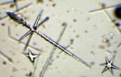
Figure 3. Microscopic views of various sponge
spicules. Left: Three of the more than 30 different
types of spicules found in demosponges. Center:
The six-rayed hexact spicules are found only in the
hexactinellid sponges. Right: Three-rayed calcareous
triact spicules are characteristic of calcareous sponges.
|
Step 2a. Identification of Demosponges to Species
If you have determined your sponge's
major group and you are a brave, or foolhardy, soul, you might
wish to try to determine its species. This may be a very difficult
task, however; rather like the old saying about exceptions
proving the rule, some demosponges ARE easy to identify. These
are sponges whose the colony has a more-or-less defined shape
and color. In tropical areas these are generally larger sponges
and are found in high current areas. As such, they are not
suitable for reef aquaria; they are either too big or they
require both high currents and laminar water flow. Although
many reef aquaria seem, at least to aquarists, to have impressive
water turn-over rates, these rates are often small compared
to the several knot currents that flow along, and over, reefs
at flood tides. Additionally, virtually all aquaria are dominated
by turbulent flow regimes, and these flow patterns are not
suitable for many sponges. The natural environment's high
water velocities and consistent laminar flow have worked as
naturally selective agents, however, and this has resulted
in the distinctive shapes of many of these sponges. These
sponges may sometimes be identified by comparison with published
images, provided, of course, that those images were correctly
identified in the first place. Generally, large and distinctive
sponges can be identified reasonably well by comparison to
photographic guides AS LONG AS THE SPECIMEN'S GEOGRAPHIC ORIGIN
IS KNOWN. Knowing the geographic locality is important, as
different species from different regions may look effectively
identical in photographs.
|
|
Figure 4. A couple of sponges that are identifiable
by comparison to photographs: the purple sponges are
Niphates ramosa and the orange sponges are a
species of Agelus. They were identified by H.
Reiswig, one of the premier workers studying sponges.
The individual Niphates colonies are over an
inch in diameter and are unsuitable for reef aquaria.
|
Sponges are found in many habitats, not just those dominated
by laminar flow and high currents. In fact, sponges are found
in just about all possible combinations of water currents
and flow types, from high current areas in surge or highly
turbulent areas to extremely calm waters. Sponges from many
of these types of areas may do quite well in aquaria. Unfortunately,
such sponges tend to mold their shapes to the currents and
substrata surrounding them. In other words, the same species
will have differing appearances in differing habitats. In
the most extreme examples of this environmental sculpting
of shape, many sponges change form and appearance as they
grow, responding to changes in the water currents generated
by either their own shapes or by nearby shapes in the environment.
Such sponges have a labile shape and are damnably difficult
to identify; about the only way of ensuring a proper identification
is by comparing a microscopic examination of the sponge's
spicules to what is known from the technical literature of
the sponges from a given area. For such a process to work
several different things must occur.
-
First, the area of the sponge's origin must be known.
-
Second, the aquarist has to have access to, and know
how to properly use, a microscope.
-
Finally, the aquarist has to have access to the technical
literature describing the sponges by spicular type.
|
Figure 5. The types of spicules found in one
species of sponge (From Kozloff, 1996). For a valid
identification of the sponge, not only do all the types
of spicules have to be present, they have to be of the
appropriate size and be present in the proper relative
abundances.
|
The odds of satisfying all of these requirements are slim
to none for most hobbyists and, because of that, many of the
common demosponges in their tanks cannot be identified with
any degree of certainty. This results in an interesting paradox;
the sponges that they can identify by comparison to photographs
are ones that generally can't be kept, and the sponges that
generally do well in tanks are not easily identified.
At this point, I should probably discuss a small group of
sponges called "Sclerosponges." These animals secrete
siliceous spicules imbedded in a calcareous matrix, and the
living cells form a layer upon and covering this stony matrix,
much like tissue frosting on a calcareous cake. Sclerosponges
are somewhat uncommon, but are generally found living on coral
reefs. They were first discovered living in caves, which is
one of their preferred habitats. Although unlikely, it is
possible that they have been collected incidentally for the
reef hobby. The only way these sponges can be identified is
by examining the rocky matrix underlying the tissue frosting
to find the silica spicules it contains. Superficially, they
appear to be a thin layer of sponge living on rock; however,
in this case, they actually secrete the "rock."
For a few years, from about 1970 to about 1985, many authorities
considered them in a separate class, but a lot of research
has showed that the "Class Sclerospongia or Stromatoporoidea"
is an artificial grouping of several superficially similar
sponges from several demosponge subgroups. Basically, this
"grouping" is a good example of convergent
evolution at work.
|
Figure 6. A sponge that is possibly a sclerosponge
photographed on the roof of a cave in a Caribbean reef.
The surface's "star-like" or "stellate"
pattern is characteristic of some sclerosponges, but
the sponge's identity must remain uncertain, as the
specimen was not collected for definitive identification.
The field of view is about 5 cm (2 inches) across.
|
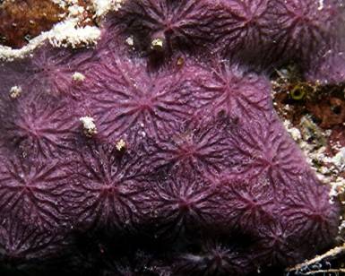 |
Step 2b. Identification of Calcareous Sponges
Compared to demosponges, the calcareous
sponges are significantly less diverse, and the ones typically
found in aquaria are relatively easy to name. Whether or not
that name is a "real" one, well, that is open to
question. Calcareous sponges are generally small; although
some species get quite large in nature, colonies in aquaria
that are an inch in size are giants. In natural environments,
larger species may be relatively common, but such species
are not often found in aquaria. The typical aquarium example
is cylindrical or tubular, and while other colors are found
in nature, aquarium species are almost uniformly white, tan
or a drab, nondescript gray.
Probably the type of calcareous sponge most likely found
in reef aquaria are the so-called "Pineapple" sponges.
These small, white or gray sponges often appear in a reef
aquarium a few weeks or months after it is set up, and may
or may not persist for a long time. They tend to appear in
areas of relatively high current flow, and big ones reach
heights of an inch or so. Aquarists commonly say that they
are in the genus Scypha. This may be true, but see
the discussion and example in the next couple of paragraphs
for the problem: calcareous sponges of essentially the same
shape, size and color are described from different areas under
the generic names of Scypha, Grantia, Sycon,
Leucilla and Leucandra. Species from these species
cannot be distinguished by cursory examination. Snap "off-the-wall"
identification by aquarists is particularly problematic with
the calcareous sponges, which tend to be smaller and more
symmetrical than most other sponges.
A Few Words of Caution
Even within a group that has consistent
structures and shapes, the identification of sponges is not
an easy task. For example, not only do some species of calcareous
sponges in the genera Scypha, Grantia, Sycon,
Leucilla and Leucandra look alike, small individuals
of species from the other classes of sponges can have the
same shapes, colors and symmetry. The ONLY way to really
tell all of these animals apart is by microscopic examination
of spicular patterns and abundances. Nevertheless, some aquarist
literature references and other "experts" claim
to be able to tell these various species apart using only
pictures or simple verbal descriptions. This is simply not
possible.
To illustrate some of the difficulties of sponge identification
even within a group of "well-characterized" and
"well-known" species, I have included the following
portion, called a couplet, of a taxonomic reference specifying
the differences between two calcareous sponge species. Several
choices precede this choice, basically determining that the
body is wrinkled, flattened and sac-like.
Choice 1:
Height typically 2-3 cm, width 1-2 cm; with a very short
fringe of oxeas around the osculum; oxeas 150-500µm,
triact rays 40-150 µm, short tetract ray 50-60 µm,
long tetract rays 60-90 µm...
Grantia ? compressa
Choice 2:
Height typically 6-16 cm, width 2-5 cm; without an evident
fringe of oxeas around the osculum; oxeas 400-500µm,
triact rays 120-350 µm, short tetract ray 40 µm,
long tetract rays 150 µm...
Leucandra similar to levis
|
Figure 7. Left: Grantia or Scypha
sp. Right: Leucandra sp. These possibly
are the sponges discussed in the taxonomic key couplets
in the preceding paragraph. Note that while individuals
of the two species may appear to be distinctive, it
would be impossible to distinguish the lower right individual
of Leucandra from the individuals of Grantia
or Scypha on the basis of appearance alone.
|
The distinction between these species is pretty clear, provided
that you can determine which spicules are called oxeas, triacts
and tetracts, as well as being able to measure them. To use
this couplet of descriptions, you need a compound microscope.
Additionally, although using this couplet to identify the
sponges seems to be straightforward, notice the many places
where the characteristics overlap - and these are not closely
related animals! Secondly, note that when finished, both choices
lead to incompletely described species. This faunal key (Kozloff,
1996) was published in 1996 and covers one of the world's
better-known marine faunas - and yet it is still impossible
to definitely identify some common sponges. The sponge fauna
of most tropical marine areas is poorly characterized and
described... and yet we have people foolishly purporting
to be able to identify sponges by pictures alone.
Aquarium Husbandry
I suppose that after "What is
it?," the next question most aquarists ask is, "How
do I keep it?" Unfortunately, for this group, as well
as for other diverse animal groups, there is no simple good
answer. There are well over 5,000 species of sponges, and
as with all other animal groups, each species is adapted for
its own particular set of conditions. There simply is no "one
size fits all" answer. Unfortunately, most aquarists
keep trying to prove that there is. A lot of animals get killed
in such endeavors.
To keep any animal in good health, it must be kept under
the environmental conditions in which it does best. In the
case of reef sponges, probably all of them will thrive under
the salinity and temperature conditions of the average tropical
coral reef; in other words, a temperature around 82°F,
with a salinity of 36 psu.
Such conditions are easy to duplicate in aquaria. And they
are a good first step.
But they are only a first step.
The primary environmental condition that will determine the
survival of any sponge is the water flow regime. To understand
why water conditions are important, it is necessary to review
how sponges gain nutrition. In general, sponges fill a volume
with "tissue" and pump water through that volume
to extract usable particulate food. A simple tubular or cylindrical
sponge with its food collection area on the inner surface
of a large cavity "wastes" a lot of internal space.
Some of this is "dead" space caused by water currents,
other space is wasted when water currents carry the suspended
particulate material too far from the choanocytes. Consequently,
most sponges' filtration area has been maximized by filling
the cavity with - dare I say it? - a "spongy" mass
of small chambers, each lined by choanocytes. Large sponges
may have more than 10,000 chambers/mm3 (164 million chambers/cubic
inch) containing, in total, many billions of choanocytes.
Some of these large sponges have been calculated to pump their
own weight of water through themselves about every five seconds.
The larger ones likely can filter several thousand liters
per day, removing up to 95% of bacteria and particulate organic
material from the water. These sponges also differ from the
"basic" sponge described in last month's article
in that their body is often asymmetrical and without ostia;
the visible pores on the outer surface are the openings of
the water channel system (Reiswig, 1975; Ruppert, et al.,
2003).
As a general rule, sponges feed primarily on very small particulate
organic material of the size category containing bacterioplankton
and the smallest phytoplankton. Larger phytoplankton also
may be eaten, but this material is generally not collected
by the choanocytes, but rather by the cell's surfaces around
the small incurrent water passages. Many tropical sponges
supplement their filter-feeding by having a symbiotic relationship
with zooxanthellae. Regardless of this symbiosis, all sponges
need to feed. They absorb dissolved organic material from
the water, but also need to have a source of bacterial or
nanoplankton. This source may be maintained in aquaria by
having a good sand bed with sufficient infauna to cause some
rapid turnover in bacterial productivity. Sponges will compete
with other bacteriovores such as tunicates and, potentially,
some SPS corals for this resource. Supplementation of the
system with small, less than 5µm in diameter, phytoplankton
cells may provide additional nutrition. Interestingly, a few
sponges that are apparently wholly carnivorous in the family
Cladorhizidae, catching prey by the action of hooked spicules
on their body's surface, have recently been described from
caves in the Mediterranean and deep seas elsewhere (Vaceletand
Boury-Esnault 1995). These animals, not found in aquaria,
eat small crustaceans.
Sponge Natural History Considerations for Aquarists
In nature, space often is a limiting
resource for marine filter-feeders. Given that sponges sit
in one place and filter water, they must have some way of
maintaining their environmental space or they would be quickly
overgrown or killed. As it turns out, sponges are masters
of chemical warfare. Some of their chemicals are potent, and
not only to marine animals. Several tropical sponges are toxic
or irritating to the touch to humans (Humann, 1992). Nevertheless,
a sponge's chemical warfare capabilities are not intentionally
directed against humans; rather, these exceptionally toxic
chemicals are meant to be effective against predators or potential
competitors for space such as corals (Porter and Targett,
1988). Because of the toxic array of chemicals they contain,
relatively few animal groups have members that eat sponges.
Some of these are the snails such as dorid nudibranchs, a
few sea stars and a few fishes (Mauzey, et al., 1968;
Bloom, 1981; Pawlik et al, 1995; Wulff, 1995). These
predators often metabolically modify sponge's chemicals for
their own defense. Both the sponges and their predators may
at times liberate copious quantities of chemically-laden mucus.
In natural situations, this chemical soup would deter predators
or competitors and then disperse. In the closed environment
of a reef aquarium, the chemicals are contained and may be
toxic to many aquarium animals. Many of these poison factories
are brightly colored, presumably to alert potential predators
of their presence. Humans tend to view these colors as pleasing
and attractive, a rather perverse twist on their presumed
purpose. Consequently, few attractive sponges or their predators
are good candidates for inclusion into a closed reef aquarium
system. Aquarists need to remember that sponges are not passive
lumps of tissue. They continuously fight for space; even if,
in your opinion, they don't need to. In this ongoing struggle,
they can and will produce very nasty substances that can affect
other animals, and one can almost never be sure which ones
will be affected and which ones will not. On the flip side
of this record (does anybody still remember records, and flip
sides?), Halichondria moorei, for example, has long
been used by New Zealand natives to aid healing. Nearly 10%
of the sponge's weight is composed of the potent anti-inflammatory
drug, potassium fluorosilicate. Unfortunately, the sponges
found in aquaria are more likely to cause inflammation rather
than heal it.
|
|
 |
|
Figure 8. Examples of sponges as competitors.
Left: The red sponge is overgrowing and killing
the coral to the left. Right: The sponge has
caused the anemone to move toward the top of the image.
Sponges often win in competitive encounters such as
these; when they do, the loser generally dies.
|
Some other sponges are not necessarily the best of reef tank
inhabitants for another reason. These sponges, the clionids,
are typically yellow or reddish sponges that are specialized
to live inside calcareous substrates. In aquaria and on reefs,
that means they erode corals away from the inside out. These
are very important bioerosive organisms on reefs, and often
what appears to be large "solid" coral heads consist
of nothing more than a veneer of coral tissue and a thin calcareous
layer over a wholly or partially eroded coral skeleton that
has been replaced by the sponge. In some aquaria, they have
become somewhat of a problem.
|
Figure 9. A coral head that is being eroded away
from the inside by the yellow clionid sponges. The sponge's
tubular osculae are visible as are "solid"
yellow masses containing incurrent water pores.
|
Some sponges may easily be kept in reef and fresh-water aquaria,
if a few precautions are followed. As a general rule, most
sponges should never be exposed to air. Many shallow water
sponges produce copious amounts of mucus, and this mucus can
form a protective layer if the sponge is exposed to air. These
species are not affected at all by even prolonged exposure
to air. Many moderate to deep water sponges, however, often
lack such a mucous coat, and if they are exposed to air, even
briefly, many of the very small passages in their water system
become filled with air and are effectively plugged. The animal
has no way to clear this air from its tiny tubules and cells
adjacent to air die, decompose and produce gases which plug
other tubes. This effect cascades and the sponge dies. These
animals are never naturally exposed to air or bubbles, and
air is deadly to them. As aquarists seldom know where their
sponges originate, it is prudent not to cause problems by
carelessly handling the animals at the air/water interface.
Additionally, it is necessary to be aware of the reef aquarium's
water conditions. If the water has been treated to remove
silica, most sponges will not grow well as they require silica
for their spicules. Calcareous sponges will do all right under
those conditions, but remember that, like corals, they depend
on the amount of calcium in the water. They also compete with
the corals for calcium, so you need to monitor the calcium
levels closely if heavy sponge growth is present.
Sponges often enter aquarium systems as incidental, and often
unnoticed, hitchhikers on live rock. If they grow and start
to spread, and you don't notice a concurrent decline in your
other animals, then you know 1) that you can keep some sponges,
and 2) the ones you have are relatively benign. Alternatively,
if you purchase a sponge, try to get one that has been maintained
in your dealer's tank for a while. Dying sponges generally
become obvious in a hurry, so if the sponge has survived for
a week or two, it may be okay. Sponges that can injure people
are unlikely to show up at dealers; however, it occasionally
happens. Ask the dealer if he has ever had a "reaction"
to the sponge. If the sponge has been kept in a tank with
other inhabitants, try to determine if those animals seem
to have reacted to the sponge. Once the sponge has been placed
in the home aquarium, monitor its condition carefully. If
it starts to develop a gray or white film growing over it,
it has started to die. Most sponges that have this film will
not recover. If you wish to try to keep the animal alive,
transfer it to a separate tank so that it will not foul your
main system's water.
Many tropical sponges have photosynthetic symbiotic algae
of a couple of types. These may be dinoflagellates similar,
but not identical, to those found in corals. Additionally,
many sponges also harbor symbiotic cyanobacteria and, actually,
quite an array of other bacteria; in some species, the bacteria
may account for as much as 40% of the weight of the sponge.
The sponge/bacteria symbioses are particularly important in
many tropical sponges - and presumably temperate ones as well
- but they just haven't been investigated there. The symbiotic
algae go a long way to providing enough sugars to fuel the
sponge's metabolism, often providing a competitive edge for
these sponges on a reef. Some of these species, as might be
expected, do well in reef aquaria, but some sponges harboring
similar species of zooxanthellae also do very well in the
deep cold water of Antarctica. One of these sponges, Rossella
racovitzae, has long (10 cm!) ossicles
that act as fiber optic light pipes to direct light
down to the zooxanthellae that live below the water sediment
interface in the mud (Cattaneo-Vietti, 1996).
|
|
Figure 10. Some hitchhiking sponges can become
pests. This calcareous sponge, similar to a species
of Leucosolenia, was sent to me by a hobbyist
for identification. The sponge has effectively taken
over most of the rock surface in this tank and has proven
essentially impossible to eradicate. Even with a specimen
and microscopic analysis, I could not identify this
species, as I did not know where the sponge originated.
|
If the animal seems to shrink or otherwise deteriorate, move
it. Some of the variables to consider with sponges are light
intensity (most species don't like really bright light - but
a few do), currents (many species like strong currents), potential
competitors (don't place it too close to either hard or soft
corals; they fight with chemicals, too) and potential predators
in your tank (keyhole limpets and angelfishes will graze on
some sponges). Once you have found a good spot and your sponge
is growing well, you will have a beautiful and interesting
addition to your system.
|


)
)
)
)
)
)
)
)
)
)
)
)
