|
Humans, in their infinite arrogance, are
prone to think of themselves as the masters of creation, and
the most important animals on the planet. This may be; but
a reasonable argument may be made for considering man as a
transitory blip on the radar screen of Earth's life forms,
and that the real movers and shakers of the world are those
animals that have multiple jointed legs and which wear their
skeleton on the outside. Such dominant animals would certainly
be the arthropods.
On land, the dominant forms of animal life,
in terms of the number of species and in terms of mass of
the animals, are certainly the insects. Literally millions
of different kinds exist, and the sheer amount of insect flesh
is almost incalculable. Arthropods dominate the oceans as
well but, interestingly enough, not the insects. For some
obscure evolutionary reason, insects are almost totally absent
from the seas. There about 30 species of oceanic sea skaters,
all in the genus Halobates, and numerous insects living
in intertidal habitats, but no insect truly lives a submerged
marine existence. This is really rather odd, as insects dominate
underwater habitats in fresh water much as they do on the
land.
Link to image of Halobates, with
information on the life history and reproduction: http://entomology.unl.edu/marine_insects/halobateslife.html
Link to information about the evolution of Halobates:
http://www.zmuc.dk/EntoWeb/Halobates/HALOBAT5.HTM
For whatever reason, the arthropod component
of the oceanic realm is crustacean. Although insects and crustaceans
shared a common ancestor, probably sometime over 400,000,000
years ago, the two groups have diverged significantly since
that time. Crustaceans and insects are very different types
of animals. Insects are conservative in their basic body plan;
they have a consistent body plan throughout the group with
little real variation compared to the crustaceans. All insects
have, for example, three body regions, three pairs of legs
on the middle body region, no appendages on the abdomen, and
internal organ systems of the same basic pattern. Crustaceans,
on the other hand, come in a very large variety of shapes,
sizes, and body forms, some having hundreds of appendages,
others having, effectively, none.
The diversity of crustacean shapes and
sizes notwithstanding, the numerically dominant animals in
the world's oceans belong to one group, the copepods. In fact,
one copepod genus, Calanus, likely contains more individual
animals over one tenth of an inch in length than any other
animal genus. At one time, it was rumored that one marine
biology professor put one of his more troublesome grad students
to work calculating the number of tons of molts produced annually
by one species, Calanus finmarchicus. The student labored
mightily and brought forth an answer of 1011 tons per year.
As each individual Calanus finmarchicus is quite small,
no larger than a small grain of rice, an annual production
of one hundred billion tons of molts per year implies huge
populations of these animals.
Links to images of Calanus and Calanoid
copepods:
http://www.ecoscope.com/copepod.htm
Calanus finmarchicus:
http://www.sams.ac.uk/dml/projects/zooplank/images/calanus.jpg
Female calanoid showing egg sac:
http://bioloc.coas.oregonstate.edu/images/photos/003x.jpg
Beautiful imagery showing appendages in colors, as well as
some great information on sensory adaptations:
http://www.pbrc.hawaii.edu/bemf/microangela/pleuro.htm
In fact, copepods are the dominant animals
in all the world's seas, and although they may be found in
densities as low one per ten or a hundred cubic meters of
water, average densities are generally considered to be on
the order of one to ten per cubic meter, and often their densities
are much greater. This doesn't sound like much until you start
to calculate the number of cubic meters of water in the oceans,
at which point you realize it sounds like a very big number,
indeed.
Copepods
Copepods are small crustaceans, generally
less than a couple of centimeters in length, in their own
taxonomic group called, not terribly surprisingly, the Class
Copepoda of the Phylum Arthropoda. There are probably close
to 7500 species with about 2000 of these being parasitic.
Some of these parasites may be quite large, for a copepod;
over a foot long, in some cases.
Unlike a crab or shrimp, copepods do not
have a carapace or "shell" covering the front part
of the body. Also, unlike crabs and shrimp, they have only
one eye, and it is on the midline of the body very near the
front end. Most other groups of aquarium crustaceans have
at least two eyes, and these eyes may (in crabs or shrimps)
or may not (in amphipods and isopods) be on stalks.
Link to an image of the front end of a
calanoid copepod showing the single pink eye in between the
bases of the first antennae: http://www.obs-banyuls.fr/Razouls/Webcd/DIAPOESP/cop5%20-%20copie.jpg
The copepod body is divided into three
parts; a front section called the "cephalosome,"
a middle section, called the "metasome," and the
posterior abdomen, sometimes called the "urosome."
As in all arthropods, the body is formed of discrete segments
or sections. In primitive arthropods, for example, such as
the brine shrimp Artemia salina, many of the body's segments
look much like the segments in front of or behind it. In advanced
arthropods, such as the crabs, there is much fusion and loss
of segmental integrity, and it can sometimes be hard to determine
where one segment begins or ends. Copepods fall somewhat in
the middle of these extremes. The cephalosome is comprised
of the five head segments that characterize all crustacea,
but also has one or two "thoracic" segments incorporated
into it. The metasome, or thorax, has four or five segments,
and the abdomen generally has four or five segments. Except
in the abdomen, each segment bears a pair of limbs.
Copepods follow the basic crustacean pattern
of limbs or appendages. From the front to the back, they have
the following sequence of appendages, one pair per basic body
segment:
In the Cephalosome:
-
The first antennae, also known as antennules.
-
The second antennae, also known simply
as antennae (if the first antennae are called antennules.
-
The mandibles, or jaws. In some predatory
Calanoids, these may be tipped with opal (amorphous silica),
presumably to harden the jaws so that they may more effectively
crush their prey.
-
The first maxillae, also known as the
maxillules,
-
The second maxillae, also known as
maxillae (if the first maxillae are called maxillules.
In the Metasome:
-
Four or five pairs of thoracic appendages,
all of more-or-less similar architecture, except in the
male, where the last pair of thoracic appendages is modified
as copulatory organs.
In the Abdomen:
|
|
|
Figure
1. Side of view of a harpacticoid copepod showing
the body regions.
|
The internal morphology of all copepods
is relatively simple. The mouth, located in front of the mandibles
on the animal's bottom surface and facing toward the rear,
is covered by a moveable flap called the labrum. The esophagus
is short and passes from the mouth forward a short distance
in the body, and then curves up toward the back and enters
the stomach. The stomach, or foregut, is relatively capacious,
and it joins to the midgut which extends through most of the
animal. The hindgut is short and found only in the last segment
or so of the body. At the juncture of the stomach and midgut
are found a pair of digestive sacs, or caeca.
Feeding and digestion in copepods is an
interesting process, and totally unlike that found in most
other animals. Movement of the food through and within the
gut is done by muscular control, not by the action of cilia,
as in many other animals. There is no mucus produced in the
gut of copepods. As food enters the gut, it is completely
encased in a thin, flexible, and permeable bag made of chitin
secreted by the foregut. This bag looks rather like a thin
cellophane sack. This bag defines, at first, a food pellet,
and then a fecal pellet. As food passes into the midgut, digestive
fluid is released, and the gut contents become acidic. There
is a wide array of enzymes together in the fluid: proteases
to breakdown proteins; lipases and esterases for fat digestion,
along with fat emulsifiers; and carbohydrases of both disaccharides
and polysaccharides, for the digestions of sugars and starches.
No chitinases are found in any arthropod, and cellulases are
absent in most of them as well. This digestive fluid bathes
the food pellet in its bag, and digested food is squeezed
out of the bag by the muscular contraction of the gut walls.
This digested food mixes with other fluids in the midgut.
Periodic contractions of the midgut force this gut fluid into
to the digestive caeca where absorption occurs. When the digestive
process is complete the food pellet is moved along to the
hindgut where it is compacted into a fecal pellet and expelled.
|
|
|
Figure
2. Harpacticoid copepod from an aquarium. Note the
red eye between the bases of the antennae. The gut is
faintly visible as a tube in the center of the animal.
|
Link to a page showing the acidity of the
midgut of a harpacticoid using a pH indicator dye. Nice photos!: http://www.sinc.sunysb.edu/Stu/miahrens/COULLANA.GIF.
The tubular digestive caeca are bathed
in blood as copepods have an open circulatory system with
a clear blood, and respiratory pigments are generally absent.
This blood carries the digested food molecules throughout
the body. The large pelagic calanoid copepods have a small
heart which pumps blood to the head, but the harpacticoid
copepods common in reef aquaria are truly heartless animals.
In these small bugs, movements of the body and gut suffice
to move the blood around.
The copepod life cycle is basic and similar
in all the groups. The adults copulate and this is often followed
by deposition of the eggs into one or two egg sacs carried
by the female. In some groups, particularly the parasitic
forms, the eggs may be shed into the sea. The eggs hatch to
release the typical crustacean larva called a nauplius. The
nauplius undergoes four or five molts, becoming larger and
adding segments and appendages with each molt. Consequently,
there are five (in harpacticoid copepods) or six (in pelagic
calanoid copepods) naupliar stages. After the last naupliar
stage, the subsequent molt remodels the animal into a juvenile
copepod, called a copedidite.
Link to an image of a copepod nauplius:
http://www.obs-banyuls.fr/Razouls/Webcd/DIAPOESP/NAUPLIUS.jpg
In this image, the gut is visible as the dark tube in the
center of the animal. The mouth is evident as the dark spot
at the top of the gut tube, in between the bases of the middle
pair of appendages (the second antennae).
Link to a different nauplius image, with the single red eye
visible: http://pantransit.reptiles.org/images/1998-08-24/Nauplius3.jpg
Copepidites have distinct segmentation,
which is lacking in the naupliar stages, and the body regions
become apparent. There are four more copepidite stages, after
which the animal molts into an adult, and becomes reproductive.
There are no further molts after this stage. For the larger
pelagic calanoid copepods, the eggs are shed in the spring,
and the first half of the life, until the first copepidite
stage, occurs in the first summer. The animal overwinters
as this juvenile stage. When the spring plankton bloom occurs,
the animal rapidly grows and passes through the remaining
molts to reach a functional adult within a few weeks. These
animals typically live about two years, although they may
live longer. In our tanks, the small harpacticoid copepods
may pass through all of these molting stages within a few
days. This short adult to adult period, coupled with the basic
high reproductive capacity of the group means that these harpacticoids
have a truly amazing capability for rapid population explosions.
Copepod Diversity
There are four distinct groups of free-living
copepods, and several types of parasitic ones. The parasitic
copepods are bizarre animals living in or on all sorts of
animals. Some of them are rather well-studied as they parasitize
salmon and other important food fish. Rarely, one or more
of these parasites makes its way into an aquarium on a wild
caught fish or within some other organism, such as a tunicate.
Those copepods parasitizing fishes are usually very evident
and easily removed. Those in other animals generally remain
unnoticed.
The four groups of free-living copepods
are the Calanoida, the Cyclopoida, the Harpacticoida, and
the Misophrioida. The last group is very uncommon and represented
by only a few species, and I will not discuss it further.
The calanoids are quite distinctive. They are generally relatively
large animals; the body may be the size of rice grain or larger.
The first antennae are very large, with more than 15 segments,
and are often highly elaborated with large bristles and other
structures, thought to increase water resistance and thus
retard the animal's sinking rate. Calanoids are exceptionally
important ecologically in that they are major components of
almost all marine food webs. Although they may be kept in
aquaria, they need specialized plankton tanks, such as plankton
kreisels, to keep them from impacting against the walls. Generally,
their culture is limited to a few progressive commercial aquaria
such as the Monterey Bay Aquarium.
Cyclopoids and Harpacticoids both may be
found in reef aquaria, but cyclopoids are not particularly
common. They may be quite difficult to distinguish from one
another. Generally harpacticoids have a smoothly tapering
body, whereas the calanoids have a pronounced constriction
just before the last thoracic segment. Often, as well, the
front end of a harpacticoid has a small pointed projection,
the rostrum, which is lacking in most cyclopoids. Cyclopoids
are typically planktonic and are very common in freshwater
ecosystems.
Link to a diagram of a Cyclopoid copepod:
http://mscserver.cox.miami.edu/MSC230/copes2.gif
Harpacticoid copepods are more commonly
benthic, living on the bottom, or epibenthic swimming just
above the bottom. Often they are referred to as demersal zooplankton,
which means pretty much the same as epibenthic zooplankton.
Demersal or epibenthic - the terms really don't matter. What
matters is that these are some of the most important food
items in natural reefs and in aquaria. They are the food of
many small-mouthed corals, small fishes, and some other benthic
animals, such as zoanthids, and small sea anemones.
In reef aquaria, harpacticoids are commonly
the first small "bugs" seen on the walls of the
aquarium shortly after it is set up. Often dense swarms of
them may occur in the water, particularly if fish have not
been added. Once the other benthic fauna starts to become
abundant, the numbers of the harpacticoids drop as predators
on them become more abundant and common. Nevertheless, unless
something becomes highly amiss in the aquarium, they never
disappear. They can be very fecund, and their life cycles
are short, often taking only a few days to go from adult through
eggs and immature forms to adult. They are major constituent
animals of the detritivore guild, and will eat small particulate
debris. Additionally they eat bacteria and microalgae that
they scrape off sand particles. In aquaria, in addition to
the adults being eaten, their eggs and larval stages are food
for small-mouthed corals such as Acropora.
While the vast majority of harpacticoids
are free-living and, in aquaria, beneficial, a few species
are found living on and, presumably, parasitizing corals.
These bugs are sometimes seen by aquarists living on some
species of small mouthed corals, such as Acropora species.
They are generally bright gold, often with a bright red patch,
and with this coloration they closely mimic some stenothoid
amphipods found on similar corals. The stenothoids are probably
harmless commensals of cnidarians in all seas, while the harpacticoids
appear to be parasitic. The significance of the similarity
of color pattern is unclear, and it really appears that virtually
no scientific research is being done with regard to either
group or their effects on the corals.
|
Figure
3. Acropora bugs. The upper right
image is a harmless stenothoid amphipod, found on Acropora.
The other three images are of a harpacticoid copepod,
probably Tegastes, which parasitizes coral. Note
the close similarity of shapes; and although distinct
differences may be seen, they are subtle. Probably the
best character to distinguish the two crustaceans is
the number of eyes. Copepods have only one, amphipods
have two. These animals are about 0.01 inch long.
|
|
|
|
|
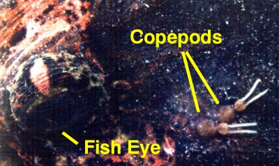
Figure
4. Blood sucking copepod parasite (Lepeophtheirus
sp.) fastened to a sculpin just behind the eye. The
white strands are egg sacs.
|
|
|
|
Figure
5. Small copepods, probably harpacticoids, found
living on a sea star, Linckia. They were about
1/32 of an inch long.
|
Conclusion:
In both the natural reef and our artificial
ones copepods are common, and important animals. The large
planktonic copepods characteristic of the open ocean are lacking
in our systems, but many of the other types of copepods are
commonly found. Their populations in our tanks may be immense;
in a large tank, the harpacticoids probably number in the
millions. Their contribution to the tank's energy and nutrient
flux is considerable and of great importance to the well being
of our aquaria. This notwithstanding, we must also be aware
that, as with most large animal groups, not all of the animals
likely to be found will be desirable. Some copepods are very
well adapted to the parasitic mode of life and these animals,
as well as their beneficial brethren, often are found in our
aquaria.
|