|
Propagation of Anchor and Brain Corals
Propagation of Brain (Lobophyllia) Corals:
Sometimes Lobophyllia corals can be separated into individual pieces without damaging any of their living tissue. Examining the colony from the underside will help you to plan the best way to separate the sections while minimizing the tissue that is damaged. If the newly fragmented sections are placed into a healthy environment, they will quickly grow new tissue over the skeleton’s damaged sections. While these corals are healing, it is particularly important to control filamentous algal growth as the skeleton’s newly exposed bare surface may otherwise become colonized, making it much more difficult for the coral to grow new tissue. If the pieces resulting from fragmentation are large enough, they may simply be placed onto the sand. If the fragments are small, it may be better to mount them onto rocks to be certain that live tissue does not become buried in the sand.
 |
|
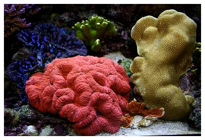
Lobophyllia can have surprisingly fast growth rates for large-polyped stony corals. I’ve found it necessary to prune two to three polyps off this particular morph of Lobophyllia about once every nine months for it to continue to fit into the tight spot in my tank. In some cases as the coral grows small areas of live tissue on the coral’s edge can become buried in the sand. These areas will die if they are buried too long. |
|
Left: Examining the Lobophyllia from the underside will help to determine the best way to separate the fragments while minimizing tissue damage. Right: In some cases the coral's skeleton is soft enough that it can be cut apart using a pair of shears, cutting pliers or tin snips. For really solid skeletal tissue, it might be necessary to use a band saw or hacksaw. |
|
Left: To be certain that the cuts with the shears go as planned, sometimes it’s advisable to pre-score the cuts with a high-speed rotary cutoff tool with a diamond-encrusted cutting wheel. Right: This particular polyp shows tissue recession on the edge due to being partially buried in sand. Under good conditions the tissue can re-grow over the skeleton in time. |
|
Left: Once separated as individual polyps, or groups of polyps, the skeleton’s base can be ground flat if the piece is too small to safely stand in the sand on its own. Right: The fragment’s base is dried prior to applying the gel cyanoacrylate glue. |
The fragments can be mounted using gel cyanoacrylate glue or putty epoxy as described in a previous article. Cyanoacrylate glue accelerant is spotted onto the substrate using a cotton swab (left), and the fragment is quickly put into position (right), being careful not to get any water on the glue's surface prior to its contact with the substrate. |
|
Shown about five weeks after the fragmentation process, this healed fragment is doing very well. The concrete mounting square is invisible beneath the surface of the sand. |
Propagating Flabello-Meandroid Euphyllia/Anchor Corals:
The propagation of branching anchor/hammer (Euphyllia paraancora) or other branching Euphyllia corals was covered in a previous article in this series. Propagation of branching Euphyllia is straightforward and entails very little risk to the coral because usually no live coral tissue need be damaged. Flabello-meandroid (corals with a continuous skeleton) Euphyllia corals, on the other hand, normally have no natural gaps in their live tissue.
As with all corals, it is best to attempt fragmenting only a healthy coral. The exception to this rule is if a coral has some type of infection that has resisted attempts at treatment, and the only way to stop the infection may be to cut out all diseased tissue. When healthy, Euphyllia corals' live tissue typically extends down onto their skeleton an inch or more from the point to where their tentacles extend.
|
This anchor or Euphyllia ancora coral has outgrown its spot in the tank and is in need of a trim. |
|
With the tentacles retracted it’s easy to see the skeleton’s pattern and plan where to make cuts. This photo clearly shows where the live tissue ends (the live tissue is brown) and the skeleton continues. |
Flabello-meandroid anchor corals can be propagated simply by cutting through the skeleton using a high speed rotary tool with a diamond-encrusted cutoff wheel (photo above left). As for Lobophyllia, as described above, the skeleton can be scored with the tool, and the sections then can be broken apart by hand or with a pair of shears, cutting pliers or tin snips (photo above right). There may be some value in cutting the coral’s live tissue with a sharp razor blade or scalpel, rather than letting it tear. Cutting the tissue may minimize the surface area of damaged live coral tissue and thereby reduce the chance of microbial infection.
|
|
After several weeks, fragments are comepletely healed and tissue has grown over the damaged skeleton. |
|
Similar to what was done above with the Lobophyllia, once the coral pieces have been separated, I like to grind a flat spot on the base and mount the coral to a piece of rock or concrete using either cyanoacrylate glue or underwater putty epoxy. To minimize the potential for a microbial infection attacking the damaged live coral tissue, it is important that the newly fragmented coral not become partially buried in the sand. Mounting the coral to a stable substrate minimizes this risk and makes it easier to handle the coral without causing additional tissue damage. It may take several weeks to a few months for live coral tissue to grow over the exposed skeleton and reform tentacles in that area. During this time it is important for the coral to be exposed to good water circulation to wash away any dying tissue. There is a balance to be maintained, though, as Euphyllia genera corals do not appreciate really strong water motion. A gentle swirling of their tentacles every few minutes is a good flow level to strive for.
I’ve also used the above technique to very successfully propagate Nemenzophyllia (fox corals) and Galaxea (galaxy/starburst corals). Fox corals' skeleton often is so fragile that the coral can be broken apart with bare hands. The techniques I’ve described here are quite likely also applicable to Catalaphyllia jardinei (elegance coral), Plerogyra (bubble coral), Physogyra (bubble coral) and innumerable genera of "brain" corals.
Happy fragging!
If you have any questions
about this article, please visit my author forum
on Reef Central.
|

