|
"In the end, we will conserve only what
we love, we will love only what we understand, and we will
understand only what we have been taught." Baba Dioum,
In Nature
Overview:
The wise prelude
above offers, I think, elegant words for a simple philosophy
that most of us practice. Some of us love what is popular,
and conformists are taught that popular is what we want. If
you ask a general question on most 'Net message boards about
what to keep in a new aquarium, or if you browse the online
and printed picture galleries in our hobby's literature, you
will see the same reef species and the same aquascape styles
over and over again. Traditional choices are so pervasive,
yet beautiful, that most of us accept these familiar organisms
and designs without exploring beyond them for newer or more
creative options. Mind you… this is not a matter of right
versus wrong or good versus bad display choices. But simply
stated, I am sure many folks would readily try alternate specimens,
designs and techniques if only they were encouraged to do
so. There is far more to the aquarium hobby than keeping the
traditional staples.
The topic of this article, as you can see, is fluorescence.
I dare say it's an issue about which most aquarists feel they
understand what "little" there is to know. Some
corals fluoresce and some don't, right? Certain light bulbs
produce more dramatic colors and "effects" of fluorescence
than others, right? Yet the truth of the matter is that what
we have been conditioned to expect from fluorescent organisms
under popular hobby lamps (blue-weighted) is merely the tiniest
fraction of what is actually going on with fluorescence! Whether
you are a casual aquarium hobbyist, a scientist or simply
an intelligent and curious reef aquarist, the world of fluorescence
is amazing and remarkable to explore with applications to
satisfy aesthetic as well as academic interests.
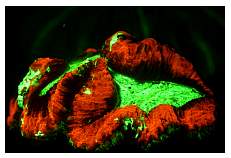
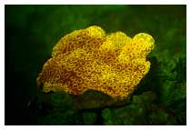
|
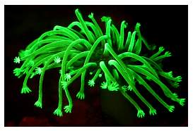
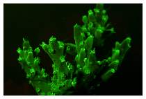
Sarcophyton: a comparison - with and without
daylight.
|
I think it is safe to say that, to many people, reefkeeping
is truly a marriage of scientific and avocational interests.
The complexity of relationships between the organisms we choose
and other life forms, their environment and the hardware that
supports them, necessarily drives us to want to learn and
observe more and more all the time. To the point, we (reef
aquarists) have both the potential and the drive to investigate
some amazing dynamics of aquatic science through our hobby.
With proper instruction we can do it with an elegant weave
of chemistry, physics and biology applied in our husbandry.
In many ways some of us are practicing amateur scientists.
And we have tremendous resources, namely livestock and disposable
income, that are a unique treasure for work in science at
large to utilize.
Quite a few professional scientists know this to be true;
we count many among our hobby's friends who eagerly work,
or otherwise interact, with the private aquarium community.
Some hobbyists had the pleasure to see Dr. Charles Mazel,
for example, present the topic of fluorescence at the 2004
IMAC conference. We were truly inspired to hear what Dr. Mazel
had to say, and I think he was impressed in kind by the knowledge
and potential of reef hobbyists that he met. The equipment
I use to study and photograph fluorescence on land and under
the sea is from his company www.nightsea.com.
Dr. Mazel has spent a lifetime on his work with coral fluorescence.
His research is fascinating, his passion for the sea apparent,
and his products are offered from a love of studying the same
organisms that reef hobbyists admire. Please be sure to explore
the resources and references of Mazel, et al that I
have cited below. They contain a lot of fascinating reading.
In this article I intend to detail some of the benefits to
aquarists of observing the fluorescence of marine organisms.
My hope is to inspire folks to explore even just a tiny bit
more of the potential for discovery, wonder and enjoyment
in the learning centers that we call reef aquariums.
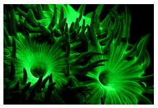
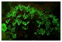
|
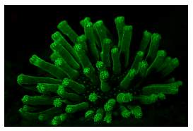
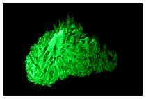
With tentacles extended or not, this Sarcophyton
makes quite an impression by night.
|
Definitions:
So what exactly is fluorescence? For
starters, not all things that luminesce in our aquaria are
fluorescing. Some organisms bioluminesce, such as the
small worms or Ophiuroid starfish that we see suddenly beaming
brightly at night when stimulated. Bioluminescence
is a product of a chemical mix that produces light. One form
of energy is converted into another. We see this in some bacteria
and among various macroscopic life forms. Some fishes and
squids house such bacteria in symbiosis (e.g., "Flashlight"
fishes). The process of bioluminescence can be intracellular
or extracellular, but its chemical origins distinguish it
from fluorescence.
Another form of light emission that we see, in minerals,
for example, is phosphorescence. Phosphorescence
is the glowing emission of light from absorbed radiation
(a source of excitation). Unlike fluorescence, though, the
glowing emission (phosphorescence) continues for a period
of time after the source of light energy has ceased.
There are other "shiny" issues, too, that we must
contend with. Some of the very colorful reef creatures have
no remarkable distinction under a light that excites fluorescence.
This is often because of still different artifacts in how
we perceive color and light… namely reflective
or iridescent qualities of various structures (spicules,
e.g.), pigments and scales.
But fluorescence, in layman's terms, is the absorption
of light at one wavelength and its re-emission at another
(without heat). Note: the difference in wavelengths (re-emission)
is an important distinction here from mere reflection.
Fluorescence changes the wavelength (color); reflection does
not. Yet not all fluorescence is apparent to the naked eye;
hence the excitement you hear from folks fascinated with the
tools used to help see and record fluorescence in reef organisms.
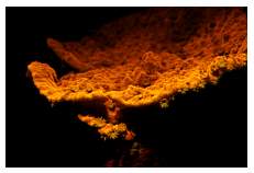
|
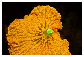
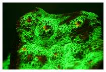
This Tongan Acanthastrea is so unremarkable by
day that you'd easily swim by its discreet brown visage.
But at night with fluorescence... it comes alive on
the reef!
|
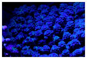
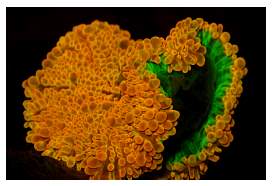
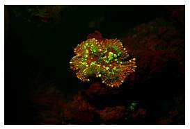
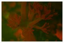
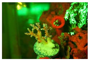
Seeing Fluorescence:
|
|
|
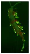
The fluorescing visible in this nudibranch is
two-fold: the slug's own green pigments, and the
red color of the algae in its 'gut' (tassels of
cerata).
|
|
Indeed, many corals
have no hint at all of (visible) fluorescence by daylight.
But seeing the "big picture" with fluorescence is
not about daylight, or even reef corals solely. Sponges, algae,
worms… even some fishes and many other organisms will
fluoresce with the right source of light and filters to see
or record it. This is one of the first things to really astound
aquarists when they begin exploring with fluorescence tools.
Everything that has been so familiar to us - diving reefs
by daylight (or night with a flashlight) and viewing our aquariums
under traditional hobby lamps - is cast in a dramatically
different light, quite literally, in the dark with
fluorescence. You feel like you are exploring an unseen new
world because, in a very real way, you are.
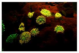
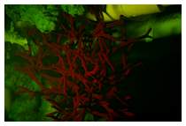
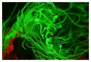
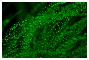
While it's true that green is the most common fluorescence
color, fluorescence explorations reveal a bounty of other
magnificent colors in red, orange, yellow and cool spectrum
colors alike. One of the very first things to impress aquarists
is… red. Red, red… crimson red, everywhere!
The reef is covered and coated in a most beautiful red color
from the fluorescence of chlorophyll (the primary light-absorbing
pigment in plants and algae). Interestingly, a number of gorgeous
Xeniids such as the blue, green or silvery pulse corals also
reveal a magnificent crimson color (photo below); it's quite
a stark contrast to what we are familiar with seeing in such
soft corals. Some feather dusters that do not appear to have
any remarkable color by day also come alive in fluorescence.
These are but a few examples.
 |
 |
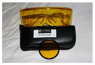
To better view fluorescence in aquaria,
we need a proper source of light (a specific excitation spectra)
to stimulate the fluorescent emissions. Special yellow glasses
will filter the extraneous light for blue light fluorescence.
You'll also want to turn off aquarium lamps and indirect room
lights. This is even more important for your camera, which
does "see" some reflected light that we cannot.
For imaging, a barrier filter is likely available for your
camera if your lens is threaded. Otherwise, get creative on
a do-it-yourself project to capture the weakly fluorescing
emissions. Thus, with glasses for viewing aquaria, or a lens
to fit over a diver's mask, and a filter for the camera and
flash… all eyes can now see the bigger fluorescence picture!
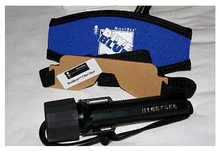
All we need now is a source of excitation (other than Rod
Stewart classics and too much wine). For this, our traditional
actinic lamps and blue spectrally weighted bulbs are useful,
but specialized excitation filters for camera flashes and
intense LED flashlights can provide a much more focused source
of excitation. Photo tip: you may need to leave a tiny
bit of lingering daylight (a weak desk lamp, for example)
or red light on in the room for your camera's lens to focus
on objects; the requirements vary by camera or lens. Best
fluorescence viewing, however, is done through the yellow
glasses/lenses in darkness with a focused source of (blue
light) excitation such as a flashlight and/or filtered camera
flash. Once the glasses, lenses/filters and lights are all
in place, the fun begins! Be prepared though… fluorescent
colors are quite vivid; it's an aspect of their very narrow
wavelength (a rather pure spectral product).
 |
 |
The tools used to assist the human eye and camera lens to
see and record fluorescence are
rather affordable, with supplies ranging from roughly $100-300.
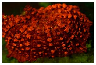
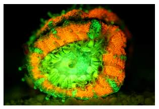
Why Fluoresce?
Coloration in reef
organisms at large is quite a complex phenomenon. Much attention
has been paid to such issues by aquarium hobbyists and researchers
alike. The matter is complicated further by the very limitations
of how different people interpret the same colors. That, however,
is a matter of psychology, to some extent.
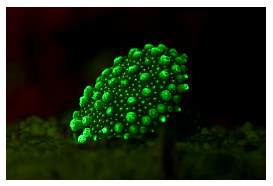
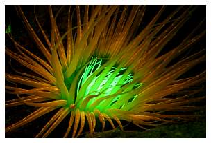
|
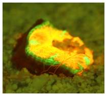
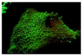
This gorgeous corallimorpharian could not be much less
attractive by daylight! Very pleasant discoveries in
a whole new realm appear when using fluorescence imaging
tools.
|
We know that corals specifically produce
proteins that fluoresce while hosting symbionts known as zooxanthellae.
The fluorescent proteins (FP) may be utilized in part to reflect
or re-emit light as needed. Whether you know it or not, most
aquarists are quite familiar with this process. But first,
let's examine the purpose of FPs.
|
|
|
Numerous organisms that might not otherwise be discovered
in the recesses of the reef, such as this tiny feather
worm, are apparent with fluorescence tools.
|
Some pigments that make corals appear colorful to our naked
eye may be used to reflect excess/"unwanted" light
away from shallow-water adapted corals. Or, if the proteins
are fluorescing they are probably not "reflecting,"
but rather the pigments absorb (and then fluoresce) to remove
the excess light. In lower light, such absorption and re-emmision
expands the spectral range of useful light (range of PAR)
for the hosted endosymbiotic
algae.
|
|
|
Notice the worms fluorescing just in front of the Acanthastrea
polyp.
|
In either capacity, the presence of FPs produces the re-emission
of sharp spectra of light that we can see as fluorescing.
So, when truly shallow-collected corals are no longer kept
under adequate light (less intense/less UV… whatever)
they may "lose color." What we are then seeing,
at least in part, is the reduction of FP density in the absence
of peak light: a significant biological savings. Similarly,
some deep(er) water corals - such as blue or green Plerogyra
corals or bright green Nemanzophyllia Fox corals -
may lose color when placed under brighter lights. They
too are reducing their density of FPs and may appear browner
colored from the now more apparent and "unmasked"
populations of (brown) zooxanthellae. These are not the only
possible reasons for various corals losing color, but they
are influences that often contribute.
Whichever, if any, of the above-listed roles FPs play - reflector
or light-harvester - the application of specific qualities
of light stimulates their production and presence. Much to
the chagrin of reef gardeners, the exact light required for
producing the beautiful range of colors seen on a reef varies
for different corals with different FP. This is but one of
the many challenges of maintaining a garden reef aquarium
under standardized parameters of light, water flow, feeding,
etc. It is also one of my favorite rants to fellow aquarists
in encouraging more folks to get away from impractical, if
not impossible, garden reef aquaria with very unnatural mixes
of specimens from different oceans or parts of the reef. I'm
encouraging friends to focus on more natural and compatible
groupings, if not specific biotopic displays. Well… it's
either that, or I'd at least like to hear less folks complaining
that all their corals are not all thriving with
all of the original colors they were purchased with…
in their homogenized garden reef display. Sheesh! It's an
unrealistic expectation.
Frequent readers of my writings might recall the popular
example that I like to use to illustrate this dilemma: Acroporids.
The family Acroporidae is… er, huge. And an aquarium
dedicated to keeping so-called SPS (small polyped stony) corals
even so "specifically" as Acroporids is really not
so specific at all. Diving the reefs reveals that the two
genera from this family that are principally used in the hobby,
Acropora and Montipora, largely contain species
that have such widely differing requirements that it is no
wonder that members of both groups in the same aquarium usually
do not fare equally well in color or growth. The optimal water
flow and lighting requirements are really quite different
among members of Acroporidae and many commonly kept (together)
corals in general.
 |
 |
Reef aquarists often realize with chagrin that the lighting
requirements of even related family members like Acropora
and Montipora are so very different as to stymie the
optimal keeping of both groups in the same aquarium.
Photos courtesy of Robie Sayan (ROBZ).
Uses for Fluorescence Imaging Tools:
One of the many
interesting uses for fluorescence technology is for studying
the early life history of settled cnidarians (Mazel, C. H.,
M. P. Strand, M. P. Lesser, M. P. Crosby, B. Coles, and A.
J. Nevis, 2003). This one has applications for coral farmers
and reef scientists alike. With specialized underwater optics
and imaging equipment, we can observe and record specific
fluorescence and reflectance patterns of rather minute or
diminished emissions. That is to say that by noting unique
occurrences of distinct emissions of light, we can spot the
very tiniest and earliest stages of cnidarian (and other fluorescing
organisms') settlement. I must admit, this was one of the
first surprises that I had when exploring my tank with NightSea
goggles and a flashlight. Not only could I spy the almost
microscopic settlement of hundreds of planulated Pocilloporid
larvae (albeit not an uncommon occurrence in aquaria) sooner
than usual, but I also noticed tiny buds of several other
corals in the display from which I had never harvested polyps!
It made me wonder if other coral polyps or planulae have been
produced regularly in the past, but were so tiny and unseen
that they were encroached upon and killed by other organisms
before they could grow large enough to be observed and collected.
Arghhhh! Those blasted so-called "reef-safe" shrimps
and crabs!
 |
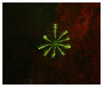
Nearly microscopic stages of settled larvae can be observed
with proper fluorescence tools long before they can
be spotted with the unassisted eye. Pocillopora damicornis,
pictured here, planulates regularly in captivity.
|
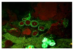
|
Some cnidarians which are less colorful than mud by
daylight, such as these brown zoanthids, are remarkably
fluorescent!
|
You can imagine just the same that researchers can scan tracts
of wild reef and interpret the data with software to get a
far more accurate assessment of species representation than
divers who tediously (and at great expense) "eyeball"
the reef to do surveys. It's a fluorescent fingerprint, so
to speak, used for mapping and assessing the world's reefs!
However, it is true that many fluorescing cnidarians share
the same or similar fluorescing characteristics. But many
fluorescent emissions are still unique and useful to observe.
Another fascinating use for fluorescence
technology is for studying the causes of coral "bleaching."
There seems to be a correlation between corals with higher
FP densities and lower incidences of photoinhibition (Hardy,
J. T., F. E. Hoge, J. K. Yungel and R. E. Dodge, 1992). Thus,
FP densities may be a reliable indicator in the identification
of key morphs of the same species that endure light stress
better than others. As we know, other environmental factors
such as temperature increases are also stressful influences
on bleaching events. Identifying the mechanisms that subsequently
force the expulsion of FP can be key in understanding some
bleaching events. The employment of fluorescent imaging technologies
is a nondestructive means of measuring such events and the
variously stressed photochemical efficiency of some zooxanthellate
organisms.
 |
 |
|
The crystal clear tentacles of this nocturnal Dendrophylliid
cup coral are nearly impossible to spot at night by
(white) flash light, yet stand out strikingly with fluorescence
in the recesses for the curious explorers.
|
Summary:
So what does it
all mean for the reef hobby? Good fodder to ponder, yes. It's
intriguing science for the more curiously minded aquarists.
Beyond the novelty and aesthetic of using such tools, we can
discover dimensions of life forms, new and unseen, in our
aquariums that we did not even realize existed. For even more
beautiful images of fluorescence, see this month's ReefSlides
here.
|
Corallimorphs that are beautiful by day can be equally
attractive when fluorescing.
|
As for the controversial issues of keeping or cultivating
select colors in corals… it really is quite a conundrum.
The practical application of light over typical reef aquaria
alone is nearly impossible to standardize. With regard for
unstable water clarity, inconsistent delivery of light (clean
or dirty paths between emission of light and the photosynthetic
creatures… namely dust, dirt and debris on lamps, lenses
and aquarium covers for starters), lamps aging and spectral
shifts, plus more than a few other influences, we may never
be able to thoroughly predict artificial reef lighting to
finesse coral coloration optimally.
Coral coloration (within reason) is not
even a very accurate if at all meaningful indicator of health.
In fact, there may be no function to some coral coloration
at all! Some theories indicate that it may all have more to
do with the "properties of the pigments, with the color
being an adaptively neutral by-product." (Mazel, C. H.,
and E. Fuchs, 2003) If so, it puts an interesting spin on
the evolution of higher order species and their coloration.
It reminds me, in fact, of a fascinating theory that Eric
Borneman was relating to me some years ago that, to summarize,
"what if" colorful reef fishes and other motile
creatures evolved their gaudy colors and patterns as more
of a means of camouflage (!) against the already gaudy and
colorful invertebrates and lower order organisms? Perhaps
adding credence to this notion is the lack of any strong evidence
that many, if any, reef fishes can see fluorescent emissions
preferentially. We must not forget, too, the sometimes-great
difference between human-perceived color and what many sighted
reef creatures are, or are believed to be, seeing.
 |
A gorgeous free-living Mussid polyp in my nano-reef.
In hindsight, though, I frankly don't care if my shrimp,
fishes or crabs agree with me on the creatures I keep and
which they think are pretty. I am attracted to this hobby
in large part for the aesthetic beauty of the creatures we
study. Fluorescence is but one beautiful dimension of that
study. Enjoy, my friends.
|
For taking pictures of fluorescence in aquaria, always
use a tripod and somewhat longer shutter speeds. Experiment
with consideration of the subject's movement, and turn
off the pump's water flow temporarily while photographing.
Also, use the strongest flash you can find with an exciter
filter to stimulate fluorescence. Use two flashes if
you can… the more excitation light available, the
better!
|
A photo tip for capturing fluorescent images: use a somewhat
faster film speed so that you can increase
the depth of field in such low light environments (darkness
is best for viewing fluorescence); perhaps as fast as
400ISO to start with for beginners.
All photos copyright Anthony
Calfo, except where otherwise noted.
* Note: if you really have some time to invest or kill…
do a general www.Google.com
search for "coral fluorescence" and note how many
hits are returned! Then, shift and refine your search with
an http://scholar.Google.com
search and see how many even still are reported: an amazing
list of content pages.
|