|
Note: Please distribute this to as
many sources, forums, clubs, magazines, stores, or other places
as possible. Please refer interested parties to this article,
or contact me by email at eborneman@uh.edu.
Background and Introduction
For many years, elegance corals (Catalaphyllia
jardinei) were among the easiest corals to keep in aquaria.
Over the past five years, most entering the trade are doomed
because of a condition for which there is no known cause or
cure. In this condition, the coral adopts a relatively swollen
oral disk with a fringe of unextended tentacles. The coral
tissue eventually shrinks, and the coral dies despite all
manner of experimental intervention.
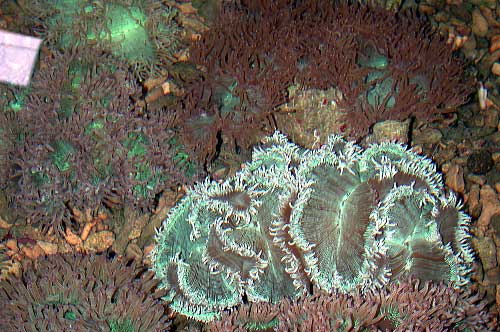
Most of the Catalaphyllia in this photo display a normal
healthy appearance,
with the exception of the one showing characteristic shrunken
tentacles
and (usually) abnormal coloration.
In some cases, a white opaque mucus-like
web may be present. I am not sure if this is an entirely separate
condition, somehow related, secondary to the primary condition,
or part of the same condition.
There has been much speculation as to why
this condition now occurs, and various sources have suggested
causes and even cures. But I stress that no research to my
knowledge has been done on this condition, and to date none
of the potential causes, solutions, or cures seems to have
much validity.
These corals are extremely beautiful and
desirable. Unfortunately today, their poor survival rate in
captivity puts them in a similar class with Goniopora stokesi
where survival rates are too low to justify the large-scale
collection of them from the wild. In fact, Catalaphyllia
appear to be relatively rare species and may be highly overcollected
so that populations in some collection areas are threatened
or even locally extinct. To continue to collect rare species
that have extremely low survival is bad for everyone - it
is an economic loss, a resource waste, and a source of great
frustration for all those who purchase and attempt to keep
them alive.
Not all Catalaphyllia shows signs
of this condition. Occasionally, I see them in stores with
a normal healthy appearance. During surveys of coral collection
areas, I never saw one with this condition in the wild, and
of hundreds being held in tanks for export, only a single
specimen showed the signs of the pathology. To be sure, Catalaphyllia
are being collected from dramatically different types of habitats,
and may be collected from very different places from where
they were collected years ago when they were easy-to-maintain.
I could speculate logically as to many potential reasons for
their current conditions and demise, but unfortunately this
speculation would be no better than the complete lack of understanding
of this condition that currently exists.
Because of the desirability and immense
popularity of Catalaphyllia, as well as to learn more
about this highly unstudied species, and to help ensure the
populations of wild elegance corals and their success in captivity,
I propose to conduct a formal study of the condition to attempt
to determine its cause and any possible solutions so that,
once again, we can enjoy healthy elegance corals in our tanks.
My research field is the investigation
of coral diseases with currently unknown etiologies. I would
like to volunteer my services to help provide answers to the
elegance coral condition. Together with collaborative work
from some of my colleagues, I believe we can determine the
cause of high mortality resulting from this condition. I will
attempt to do this in the most economical and efficacious
manner possible, and will provide results to all applicable
forums upon its completion.
Methods
I propose to collect funding and material
to conduct this work, and to do so in phases so as not to
require more funding or material than is necessary.
Catalaphyllia will need to be acquired
from various sources, both healthy and affected with the condition.
In some cases, special shipping arrangements might be required
to avoid delays or exposures that might confound any pre-exisiting
conditions from the wild. Corals will be sent either live,
or dead and preserved for analyses. Generally, a type of formalin
or alcohol fixative will be used, and not everyone will have
access to some of the fixative material. I will then examine
the corals working from the most obvious to the less obvious.
External and gross changes will be documented in live samples
with a clear description of the signs, changes, time frames
and fate of the coral over which the condition occurs. I will
look at the colonization of the surface flora and fauna by
using sterile swabs and take samples for live material and
freeze them for any future molecular work that might be required.
I will prepare histoslides to examine microstructure and look
for obvious abnormalities in tissue or zooxanthellae and the
presence or absence of intracellular parasites or pathogens.
At this point, something may or may not make itself apparent.
I will prepare a report, offer my best suggestions for the
next phase if the cause is still unknown, and outline the
next funding and materials request.
Initial Requirements
Initially, at least, the only thing I would
need is corals - as diverse in time and space as possible.
For example, ordering 100 corals from the same wholesaler
on the same date would be far less valuable than a handful
from a number of sources over various time periods to increase
the sample diversity. I will need elegance corals from as
many sources as possible. The following are sources that should
ideally be exploited for sample material.
Direct from the wild
Export facilities
Wholesale facilities
Retail facilities
Home aquaria
Healthy Corals - The Hard Part
Live corals
I would like to obtain 5 -10 healthy elegance corals with
at least two of these arranged to be shipped directly from
the source to my lab, or to a facility who is willing to ensure
that the specimens go directly into a clean covered tank with
freshly made seawater using bottled distilled water and clean
gloved hands. See protocols below. I will be attempting to
contact sources for this material, although if anyone has
such contacts and abilities, their efforts would be greatly
appreciated.
As for the remainder of the healthy samples,
it would require someone with enough experience to determine
if a given specimen truly appears to be one unaffected by
the condition. This requires either a facility with a regular
turnover of Catalaphyllia or volunteers policing local
stores to find the rare ones that are healthy. In any case,
I think a photo should be sent to me prior to acquisition
or shipping for confirmation that it does appear to not be
affected. If it turns out to be affected, it's not a problem
as it will still contribute to the study. However, I have
to acquire live healthy specimens as a control. If a healthy
(or suspected to be healthy) coral is located and provided,
I need that coral to be carefully collected and shipped to
avoid any new sources of contamination. The process will be
described below in the "techniques" section.
Tissue samples
I know I have seen at least several photos from aquarists'
tanks who have apparently healthy elegance coral in their
tanks. I have one, as well. For those who have healthy elegance
corals and want to keep them in their tanks (likely everyone!),
a tissue sample can be used. The techniques section below
outline the procedure.
Diseased Corals
Live corals
Catalaphyllia showing signs of the condition can be
prepared and sent using the same protocol for healthy corals.
These can be from any source where they are found.
It is very important to ensure that no
additional contamination occurs until I receive them. For
example, if this condition is caused by a bacterium, and aquarists
grab the coral with their hands and I run surface samples
and find most or all have the unusual presence of non-marine
types of staphylococci on them, I would erroneously look at
the staph bacteria as a possible causative agent. It would
be wasteful to run culture and reinfection studies looking
at fulfilling Koch's postulates when these microbes were all
artifacts of human handling. The same is true of air exposure
- we really want to limit the number of mold, fungus, bacteria,
and other microbial organisms that are common in the air and
on surfaces that could confound the tests and make the study
longer, more expensive, and more difficult.
Fixed dead corals
If an affected Catalaphyllia is found and cannot be
shipped live to me, I can use the whole coral or tissue samples
of fixed material. For example, if someone has purchased or
seen a Catalaphyllia that they feel has no chance for
survival, and might not even last through the shipping process,
it can be killed and fixed for study. Similarly, stores and
aquarists with access to affected corals that are willing
to donate tissue samples or the corals, but only if they are
not alive for whatever reason, can do so through fixation.
The techniques section below
outlines this procedure.
Skeletons, Photography, Direct Counts, and Surface
Swabs
If anyone wants to contribute to this study
but cannot contribute in any of the ways above, there are
still some possibly important bits of data that could be obtained.
I can use skeletons of dead Catalaphyllia, photos of
affected living Catalaphyllia and photos of the skeletons
of recently dead Catalaphyllia, and sterile surface
swabs of affected corals from tanks or from facilities. None
of these do I feel will provide conclusive evidence of anything,
but could potentially be used to support other findings.
Skeletons can be valuable because of the
presence or absence of various organisms. The presence or
absence of boring algae, fungi, mollusks, crustaceans, and
other flora and fauna may be occasionally or consistently
present and might deserve further investigation as to whether
they play a role in the condition.
Photography is supportive and may be good
in terms of documentation. It may also be able to provide
evidence of gross changes or factors involved in the condition.
Another bit of information that would
be helpful is epizootiological. It would be good to have some
idea of the occurrence of this condition. If anyone is visiting
a facility such as a fish store, wholesaler, or other source,
a simple count of the number of elegance corals present and
the number of affected colonies would be very helpful. Please
provide the date of the count, your name and contact information,
and the name of the facility, along with the count information.
You can email me the information at eborneman@uh.edu.
Sterile swabs of coral surfaces could be
valuable if the condition involves changes to normal biotic
flora, or if there is a parasite or pathogenic microbe that
effects the coral by colonizing its surface. If this is the
case, sterile swabs could be supportive if detailed tissue
work shows this to be the cause.
All protocols for this work are outlined
below in the techniques section below.
Techniques
To provide live corals to the study
(healthy or diseased)
The water in which the coral resides or
freshly-made seawater should be made using bottled distilled
water and clean salt scoops from closed containers of salt.
The coral or any tools coming in contact with the water or
the coral should not be used unless previously treated aseptically
using alcohol or a commercial aseptic scrub like Hibiclens.
To make or collect water, use a glass container
that has been wiped down thoroughly with rubbing alcohol (preferably
90%) or ethanol. All tools used to add salt and stir the container
should also be cleaned. Hands should be gloved with latex
exam gloves wiped with alcohol or hands should be washed with
an aseptic scrub like Hibiclens or wiped with alcohol. Once
washed, avoid touching unwashed skin, hair, clothing, or other
surfaces.
Water should then be poured into the shipping
bag or container. If a bag (Ziploc or fish bag), the bag should
be new and relatively sealed and having not been left open
to the air. If a container (Tupperware or similar), the container
should have its inside wiped with alcohol prior to adding
the water.
Corals should be quickly removed from the
tank and placed into the prepared water which is in the shipping
container. The tissue itself should be handled as little as
possible, grabbing the skeleton instead. For aquarists who
need to use a local store for bagging and shipping, the closed
container should be taken to the store and the same procedure
repeated for transfer to another shipping bag. Ideally, corals
will be shipped using a double bag with oxygen, with more
air than water in the shipping bag. If compressed air is used,
the filling tip should be wiped with alcohol prior to inserting
it in the bag for filling.
The container should then be quickly sealed
and shipped. All of these procedures should be timed carefully
to minimize shipping durations. Generally, this means working
in the afternoon for a later afternoon pickup. See shipping
protocol below.
Tissue sample collection
If tissue samples or fixed coral specimens
are being provided, please follow the same aseptic techniques
described above, even if it is a dead coral or skeleton. Tissue
samples on Catalaphyllia can be obtained easily by
using a scalpel, razor blade, or sharp scissors. Tissue samples
usually require some holding or handling of the coral, so
please make sure gloves or aseptically cleansed hands are
used. Once the tissue sample is obtained it should be immediately
placed into the fixative and sealed. The same would be true
of a whole colony that is affected. The colony would simply
be dropped into the fixative. I will take care of all subsequent
steps once I receive it. The amount of tissue required should
be at least 1cm x 1cm and incorporate tentacle and oral disk,
if possible and in the more affected parts of the coral. Please
do not send tissue samples without fixation. They will deteriorate
rapidly in seawater and be unusable for study. See fixative
instruction for methods.
Photography
I will gratefully accept any photographic
documentation of Catalaphyllia with the diseased condition.
It might help to just take a digital camera to a few fish
stores and snap some photos. I am especially grateful for
photographic support of corals that are either sampled or
sent to me alive or fixed. In fact, I would be very pleased
to receive photos prior to receiving coral material to ensure
that it is the right condition. I will also appreciate any
additional photos of the tank as a whole, especially in relationship
to the elegance coral. If anyone has lost an elegance coral
to this condition, but has retained the skeleton, I would
like to see photos of it from all angles and extremely close-up,
if the camera has this capability. Please do not provide photos
that are out-of focus or do not show clearly the structures
or animals being photographed. You can email digital or scanned
images to the email provided above. I do not have any practical
limitation on size of files, and I have very fast internet
lines at home and work, so don't hold back on file size or
number of files. Please provide the following information
with each photo:
Your name
Date taken
Basic photo description
Contact information
If the photo accompanies other material being sent or is a
stand-alone photo documentation.
I will probably ask other questions upon
receiving them, but this information is fine for the time
being.
Shipping
Live corals
Please use an overnight delivery service, and try to arrange
for the latest possible pick-up time so that the coral spends
as little time in transit as possible. Before you send anything,
confirm with me the planned ship dates so I can be sure to
be available or have someone available. Do not send any live
animals to my lab address as there could be significant delays
before they make it to my lab.
Send all live shipments to my home. Please
contact me by email to eborneman@uh.edu
for my home shipping address. You do not need to require a
signature release. All the drivers know me.
After confirmation with me for receiving
a shipment, please email me the tracking number to eborneman@uh.edu.
Live corals should be packed in bags or
containers and ensured that they do not leak. Double bagging
or sealing of containers is highly recommended. Make sure
that the boxes are well insulated with peanuts or Styrofoam,
and if you are shipping in cold weather, to make sure to use
a heat pack. A fish store will likely be able to provide these
and the proper number for the climate at the time. Generally,
one or two will be fine for small boxes. If the weather is
extremely cold (or hot) please wait until more moderate conditions
occur.
Fixed tissue samples, skeletons, and
other non-living material
Please send these by regular ground service
of your choice to the following address and make sure that
if liquids are present, especially alcohol or other fixatives,
that containers are rigid and well sealed, preferably with
screw-on lids rather than snap-on lids. If you are sending
glass containers, make sure the box is well packed and padded
to avoid breakage.
Eric Borneman
Department of Biology
University of Houston
Science and Research Building II
4800 Calhoun Rd.
Houston, TX 77204
Ph (713) 743-2667
Fixation instructions
I have pasted a bit of text on fixation
below from Histotechnique for information purposes. The purpose
of fixation is to preserve tissues permanently in as life-like
a state as possible. Fixation should be carried out as soon
as possible after removal of the tissues or soon after death
to prevent autolysis. There is no perfect fixative, though
formaldehyde comes the closest. Therefore, a variety of fixatives
are available for use, depending on the type of tissue present
and features to be demonstrated. There are common usages for
fixatives in the pathology laboratory based upon the nature
of the fixatives, the type of tissue, and the histologic details
to be demonstrated.
Formalin is used for all routine tissues
when an H and E slide is to be produced. Formalin is the most
forgiving of all fixatives when conditions are not ideal,
and there is no tissue that it will harm significantly.
There are two major groups of fixatives
for the work required in this project, classified according
to mechanism of action:
Aldehydes include formaldehyde (formalin)
and glutaraldehyde. Tissue is fixed by cross-linkages formed
in the proteins, particularly between lysine residues. This
cross-linkage does not harm the structure of proteins greatly,
so that antigenicity is not lost. Formalin penetrates tissue
well, but is relatively slow. The standard solution is 10%
neutral buffered formalin, although for coral tissues, 10%
formalin in seawater will work. There are a number of available
fixatives that are better, including a modified Helley's solution,
paraformaledhyde recipes and a brand called Z-fix, that are
especially good for coral tissue. If anyone has access to
these chemicals, let me know via email because this would
be the best for most of the histology assays I will use. I
can provide detailed instructions if this is something you
can do.
Glutaraldehyde fixes very quickly so is
good for electron microscopy. It penetrates very poorly, but
gives best overall cytoplasmic and nuclear detail. The standard
solution is a 2% buffered glutaraldehyde. I do not expect
to have to do EM at this point, although I might in the future.
However, I can use tissue from affected live corals and really
don't want anyone unskilled to use glutaraldehyde. It is pretty
nasty stuff.
Alcohols, including methyl alcohol (methanol)
and ethyl alcohol (ethanol), are protein denaturants and are
not used routinely for tissues because they cause too much
brittleness and hardness. Alcohols, specifically ethanol,
are used primarily for cytologic smears. Ethanol (95%) is
fast and cheap. Since smears are only a cell or so thick,
there is no great problem from shrinkage, and since smears
are not sectioned, there is no problem from induced brittleness.
Because coral tissue is only two cell layers thick, alcohol
works. It is difficult to work with the tissue later on, and
some assays cannot be performed if alcohol is used as a fixative,
but I will be able to get some results if they are used. They
are the most easily available for most persons. Ideally, ethanol
should be used by purchasing small bottles of pure grain alcohol
from a liquor store and diluting it to 70% with freshly made
artificial seawater. I would use about 1/4 of a teaspoon of
sodium bicarbonate (baking soda) in the mix, as well, to act
as a buffer, if the fixative volume is around 250-500ml (8-16
ounces).
The volume of fixative is important. There
should be a 10:1 ratio of fixative to tissue. Because corals
have skeletons, this must be included since the fixative will
soak into porous skeleton and be unavailable to penetrate
coral tissue.
Sources for materials
90% rubbing (isopropyl) alcohol for surface
sterilization is available at most major drugstores.
70% rubbing (isopropyl) alcohol for fixation
and less effective surface sterilization is available at drugstores,
supermarkets, convenience stores, etc.
99-100% ethanol is available as pure grain
alchohol from liquor stores. It is often sold under the name
Everclear. Ethanol is worth the effort to acquire, though
it costs more than rubbing alcohol. It is an extremely good
surface sterilizer, evaporates very quickly, is much more
ideal for fixation of tissues, and is relatively non-toxic
to humans and organisms in the aquarium.
Sterile swabs, syringes and containers
are available at some drugstores and laboratory supply houses.
Most veterinarians or your physician will likely be willing
to part with some, too, if you ask them.
Please contact me by email for further
inquiries.
Support for the study
If you cannot contribute coral material
but want to support the study by helping to pay for the costs
involved in the study of the condition, you may make donations
to the Elegance Coral Project Fund. Please send donations
in the form of check, money order, or electronic payment to
the following address. Funds will be maintained and used as
needed by drawing from the account. Any unused funds over
10% of the donation total will be refunded if the research
is completed without using all the funding. Refunding 10%
of donation totals will be cost and labor prohibitive, and
this remaining money will either be saved for any future projects
or donated to the investigator for his efforts in the project,
to be designated by the request of the donating party.
I am currently working on establishing
the fund, and will post the information shortly in The
Coral Forum.
Expected costs
The costs of this initial phase of the
project will be directly related to the amount of sample material
obtained. Materials costs may vary depending on what is already
available to the party. My estimates of average costs per
sample are:
|
1. Cost of obtaining the coral if
it is purchased
|
|
|
2. Materials required to collect
and ship the coral
|
|
|
3. Shipping costs
|
|
|
4. Aquarium facilities for gross
etiological description
|
|
|
5. Chemicals and disposable supplies
for sample preparation
|
|
|
6. Histological services
(2.00 per slide, 6-10 sections stained and unstained)
|
|
|
7. Prepared sample shipping costs
|
|
|
8. Laboratory supplies and equipment
for analyses of samples (may be highly variable depending
on what is found after microscopic examination)
|
|
|
9. Fees for additional researcher
expertise/consultation
|
|
It is conceivable, though very unlikely,
that the per sample cost if a coral is provided for free,
all materials and shipping are available or free, and that
the cause of this condition is relatively obvious, that the
project could be completed for the cost of the aquarium facilities
(unless donated) plus $500.00.
I suspect this initial phase of the project,
hopefully successful and conclusive, and excluding the costs
associated with obtaining the material for a good sample size
of 50 corals, will cost around $3500.00. I think this would
be a reasonable goal to try and reach in terms of initial
funding.
Absolutely Required Information on All Contributions
(except
financial only, which is voluntary)
-
Contact information including name,
address, email (phone number optional but highly recommended
if I need to talk with you quickly)
-
Tank Information
-
Size
-
Equipment used
-
Maintenance routine, including
water changes and salt brand, and any major changes
in brands or routines implemented during the ownership
of the affected coral.
-
Additives, including food, supplements,
and medications, and any major changes in brands or
routines implemented during the ownership of the affected
coral.
-
Water parameters, including but
not limited to pH, alkalinity, ammonia, nitrate, phosphate,
calcium, temperature, salinity or specific gravity,
and any major changes in brands or routines implemented
during the ownership of the affected coral.
-
Length of time the tank has been
set-up.
-
Any unusual problems or events
that may have contributed to this condition.
-
Coral Information
-
How long the coral has been in
the current tank (date of acquisition).
-
If alive, how long did it take
before signs of the condition were noticed?
-
If dead, how long did the coral
live before it died? Please include the length of
time it appeared healthy, and the length of time it
looked diseased.
-
Location where it was purchased
or obtained.
-
A written description of the condition
and any accompanying photographs.
-
Any changes in the signs of the
condition over the time the coral was in the tank.
-
Any other background information
on the coral that is known (Fiji, Indonesia, in three
other tanks before the current one, was dropped on
the floor by accident, etc.).
-
If this is not the first Catalaphyllia
owned, please provide the information above for others
and their fate.
-
The date when the coral was removed
and sent in for this study.
Example:
Joe Aquarist
15 Griggs Street
Houston, TX 77252
(713) 555-1111
email: jaqua@earthlink.net
- 75 gallon tank
- 2 years 7 months in operation
- 4 MaxiJet 1200 powerheads, 20 gallon sump,
ETS Gemini skimmer, Mag 5 return, 2 x 175 watt 6500K metal
halide, 4" CaribSea oolitic sand bed, 65 lbs. live rock,
Red Sea ozonizer, 1 liter ESV carbon changed every six months
- Kent Calcium 100 ml/week
- Kent Strontium 20 ml/week
- Brine shrimp - approx. 1 tablespoon per
day
- DT's phytoplankton, 50 ml/week
- Red Slime Remover used once, 50 mg, 10/2003
- 10% water change every week using Instant
Ocean salt. Used Reef Crystals for the first year.
- pH stable 8.0-8.2
- Alkalinity stable at 3.5 meq/l
- Calcium varies between 350-450ppm
- Ammonia unmeasurable since week 4 of the
tank
- Nitrate was 5.0 ppm but has been 1.0ppm
for past three months
- Phosphate levels stable at 0.50 ppm
- Specific gravity always maintained at
1.025
- Temperature varies: 78°F in winter,
84°F in summer
Notes: this coral was stung by an anemone six months ago,
and bleached four months ago. The condition appeared prior
to either of these events. I also added a lot of sand to the
tank three months ago and lost three other corals at the time.
This elegance coral was acquired from
Joe's Fish Store in Memphis, Tennessee in June, 2003. It looked
normal when I bought it, but developed a swollen disk and
shrunken tentacles two weeks after I put it in the tank. I
dipped it in freshwater, but it didn't change the conditions.
I tried treating it with Maracyn, but again no effect. In
August, 2003 the entire coral appeared shrunken, and a white
web formed on the surface about two weeks after it began to
shrink. The coral died in the first week of September, 2003.
I purchased another one from Online Corals, Inc. (www.sickcorals.com)
two weeks ago, and it arrived with the swollen condition.
They told me this coral came from an exporter in Jakarta.
It is still alive and its appearance is unchanged. I am sending
this coral alive to you according to the information provided
on the project page. I have emailed you photos of the coral
taken on January 25, 2004 and provided the information requested.
I will remove and send this coral to you on February 6, 2004
as per instructions.
|